|
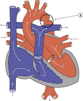
Coarctation of the aortaIn the normal heart, blood flows to the body through the aorta, which connects to the left ventricle and arches over the top of the heart. In coarctation of the aorta, the aorta is pinched (in medical terms, coarcted) at a point somewhere along its length. This pinching restricts blood flow from the heart to the rest of the body. Often, the baby also has other heart defects, such as a narrowing of the aortic arch (in medical terms, transverse aortic arch hypoplasia).
Usually no symptoms are apparent at birth, but can develop within a week of birth with the closure of the ductus arteriosus. The possible consequences of this condition are poor feeding, an enlarged heart, an enlarged liver and congestive heart failure. Blood pressure also increases above the constriction. Timing of surgery depends on the degree of coarctation. Surgery may be delayed with mild coarctation (which sometimes is not detected until adolescence). However, if a baby develops congestive heart failure or high blood pressure, early surgery is usually required. A baby with severe coarctation should have early surgery to prevent long-term high blood pressure. The coarctation can be repaired in a number of ways without opening the heart. The surgeon can remove the narrowed part of the aorta and sew the ends together, thereby creating an anastomosis. (An anastomosis is the surgical formation of a connection between two normally distinct structures.) Alternatively, the surgeon can cut the narrowed section open and sew a patch over the opening.
|
| Current | Home - Table of Contents - Chapter 2 - Coarctation of the aorta |
| Next | Interrupted aortic arch |
| Previous | Atrioventricular canal defect |
| Section 1 | Chapter 1 - Introduction to the Issues |
| Chapter 2 - Pediatric Cardiac Issues | |
| Chapter 3 - The Diagnosis of Pediatric Heart Defects and their Surgical Treatment | |
| Chapter 4 - The Health Sciences Centre | |
| Section 2 | Chapter 5 - Pediatric Cardiac Surgery in Winnipeg 1950-1993 |
| Chapter 6 - The Restart of Pediatric Cardiac Surgery in 1994 January 1, 1994 to May 17, 1994 |
|
| Chapter 7 - The Slowdown May 17 to September 1994 |
|
| Chapter 8 - Events Leading to the Suspension of the Program September 7, 1994 to December 23, 1994 |
|
| Chapter 9 - 1995 - The Aftermath of the Shutdown January to March, 1995 |
|
| Section 3 | Chapter 10 - Findings and Recommendations |
| Appendix 1 - Glossary of terms used in this report | |
| Appendix 2 - Parties to the Proceedings and counsel | |
| Appendix 3 - List of witnesses and dates of testimony | |
| Diagrams | |
| Tables | |