|
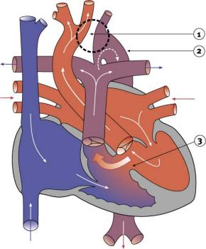
Interrupted aortic archIn this condition, the aorta does not develop completely in the area of the arch. As a result, the aorta is divided into two parts that are not connected to each other. This prevents the flow of blood through the aorta. Because blood flow through the aorta is blocked, the blood supply to the lower body is compromised. At birth, the open ductus arteriosus allows blood to flow from the right ventricle to the lower body. There are often other coexisting heart defects, such as a VSD. Interrupted aortic arch is usually diagnosed when the ductus arteriosus begins to close. With decreasing circulation to the lower body, serious damage can occur to the liver, kidneys and intestines. Prostaglandin is given to keep the ductus arteriosus open until surgery is performed, usually as soon after birth as possible. Most often a one-stage repair is undertaken, patching the arch or connecting the two ends of the aortic arch together, closing any VSD, and tying off and dividing the ductus arteriosus into two parts.
|
| Current | Home - Table of Contents - Chapter 2 - Interrupted aortic arch |
| Next | Pulmonary stenosis and aortic stenosis |
| Previous | Coarctation of the aorta |
| Section 1 | Chapter 1 - Introduction to the Issues |
| Chapter 2 - Pediatric Cardiac Issues | |
| Chapter 3 - The Diagnosis of Pediatric Heart Defects and their Surgical Treatment | |
| Chapter 4 - The Health Sciences Centre | |
| Section 2 | Chapter 5 - Pediatric Cardiac Surgery in Winnipeg 1950-1993 |
| Chapter 6 - The Restart of Pediatric Cardiac Surgery in 1994 January 1, 1994 to May 17, 1994 |
|
| Chapter 7 - The Slowdown May 17 to September 1994 |
|
| Chapter 8 - Events Leading to the Suspension of the Program September 7, 1994 to December 23, 1994 |
|
| Chapter 9 - 1995 - The Aftermath of the Shutdown January to March, 1995 |
|
| Section 3 | Chapter 10 - Findings and Recommendations |
| Appendix 1 - Glossary of terms used in this report | |
| Appendix 2 - Parties to the Proceedings and counsel | |
| Appendix 3 - List of witnesses and dates of testimony | |
| Diagrams | |
| Tables | |