|
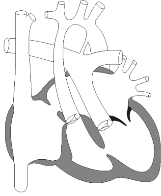
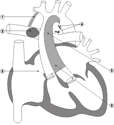
The operation-October 4In the early hours of October 4, Erica underwent a modified Norwood Stage I procedure that included
Consulting witnesses concluded that this was the appropriate repair for a child in Erica's condition. The operating team is set out in the accompanying chart.
In his report, Cornel commented that, at one hour and forty minutes, the period of TCA was very long. Odim was questioned about the length of the procedure. He gave the following response: That it was long, and as I said, we were putting this homograft. Many a times in the hypoplastic left heart syndromes, you have varying degrees of smallness of the aorta, and this child had a completely diminutive aorta due to the arch, and distally there was a coarct. So the entire area was two to three millimeters and had to be filleted open in the back, and then we had to rebuild that. The tissues were friable. The child had been on inotropes for two or three days and there was a lot of edema in the tissues. So from the technical point of view, it was a difficult type of a Norwood, given the anatomy. (Evidence, pages 25,669-25,670)
Erica's myocardial protection included deep (or profound) hypothermia with cardiac arrest. Hypothermia is a reduction in the central or core temperature of the body below 36 degrees Celsius and is achieved by infusing cold liquids intravenously and packing ice around parts of the body. With deep hypothermia, the patient is intentionally cooled to about 16 to 20 degrees Celsius. This allows the surgeon to stop the heart for about an hour and operate without either the heart moving, or tubing or blood being in the very small operative field. In his operative report Odim indicated that "30 cc per kgm. of cold cardioplegia was administered through the arterial perfusing cannula." (Exhibit 3, page BIC 56) However, there was no record in the perfusion sheets of any cardioplegia being given. Odim was questioned about this discrepancy. I can't resolve it. It is usually my practice with Norwoods to give a dose of cardioplegia. It is certainly possible that that dose was not given or it was aborted at some point, because the aorta was very small in this child. But it is my usual practice to give a single dose of cardioplegia in the Norwood setting. That practice is actually controversial. Some people do not, but I usually do, and I thought I had given cardioplegia. Q: Now, the practice is controversial, maybe you can just elaborate on that? A: It is controversial because there has been no clear benefit of cardioplegia in the setting of Norwood operations, and the use of deep hypothermia and circulatory arrest. Some people have seen a benefit, others have not. Furthermore, because the aorta is very thin and friable and small, the technique of actually trying to get it into the small aorta and into the coronary arteries is fraught with causing problems. And so for technical reasons, some people rely purely on a deep hypothermia for myocardial protection. (Evidence, pages 25,667-25,668) Perfusionist Todd Koga testified that, according to the perfusion record, no cardioplegia was given for this operation; nor could he remember any being given. He said that the manner in which Odim would have had to give the cardioplegia (through the arterial perfusion cannula) in such a case would be unusual. According to Koga's testimony: Normally it's administered from the [root] of the aorta via separate, not via the main perfusing cannula but via separate cardioplegia needle, and it is usually administered with the cross-clamp in place. (Evidence, page 7,231) Koga said that he never worked with Odim when cardioplegia was delivered through the arterial perfusion cannula. In his report, Cornel wrote: Cardioplegia was not given. I do not always use cardioplegia with PHCA [profound hypothermia with cardiac arrest] but usually do so and with an already compromised myocardium would be more inclined to do so. This is a comment rather than a criticism. (Exhibit 353, page 56) In their joint report, Cornel and Duncan wrote: We note that only hypothermia was used. However, hypothermia alone may have been a reasonable choice. (Exhibit 354, page 12) In his testimony, Cornel said: In the Norwood operation specifically, administration of cardioplegia is particularly difficult. It would be very unwise to try and put a cannula into the tiny little ascending aorta in a true hypoplastic left heart. Those vessels are difficult enough without any added trauma, and in those cases I would not give cardioplegia. In a case like this where there is access to the coronary arteries through the aorta, if cooling was proceeding very easily, I was sure that the heart was stable before surgery-I might not give it if I was a little concerned about the aortic tissue, but in a case like this I might have given cardioplegia through the aortic root with a small needle. I might or might not, but it would certainly be something to consider. (Evidence, pages 44,871-44,872) Taylor was asked if he could determine if Erica had been given cardioplegia during the course of surgery. He responded that he would have expected extensive damage to her heart if she had not received cardioplegia. He went on to say: I can only infer from the presence or absence of myocardial injury that there may or may not have been adequate myocardial protection. So I can't say, yes, cardioplegia was given or, no, it wasn't given. All I can only say is, well, she has got a lot of damage, therefore, her protection might not have been optimal. (Evidence, pages 43,234-43,235) In his evidence, Taylor also said that, while the heart was damaged, he could not be certain as to how much of the injury was sustained before the operation.
The failure to wean from bypass After the repairs had been completed, Erica was taken off bypass. Shortly thereafter, the pressure in her atrium fell rapidly and Erica's heart stopped. This necessitated a rapid return to bypass. She remained on bypass for another 15 to 20 minutes. During that time, the team adjusted some of the inotropic medication with which Erica was being treated, and attempted to go off bypass a second time. This time they were off bypass for approximately 40 minutes. However, her condition deteriorated once more and they returned to bypass. Again the inotropic medication was adjusted and another attempt was made to come off bypass. According to Odim, one of the walls (the anterolateral wall) of the left ventricle was not performing adequately. There was a progressive decline in cardiac function, and eventually Erica lost any heart rate or rhythm. At this point, the team had to decide whether to stop treatment or put Erica on ECMO. Reimer testified: We were dealing with a child whose heart was already failing before the procedure began. It was now continuing to fail, and it was just thought that the chance of her recovering, or her heart recovering, given a prolonged period of ECMO, was quite small. I mean, the risks of instituting ECMO are fairly high in terms of bleeding complications,neurologic complications and so on. And based on things like that, and I think Jonah mentioned something about the experience in other centres with ECMO and children with this, having had this procedure was not good. (Evidence, page 19,074) After consultation among Odim, Reimer, Ward, Giddins and a neonatologist, a decision was made that ECMO treatment would not be successful. Erica died in the operating room at 1630 hours. The Bichels had been advised to stay at home on the day of surgery. They received hourly updates from Lois Hawkins throughout the course of the operation. Each time she called, she told them that Erica was doing well. They finally received a call saying the operation had gone well to that point and were summoned to the HSC. When the Bichels arrived at the HSC, they were told that Erica was still being taken off bypass. They were encouraged to go for a meal. When they returned to the NICU, they were told that Erica had died. They were then taken to a private family room. At Judith Bichel's request, Erica's body was brought to the family. The parents then spoke with Odim, Giddins, Ward and Hawkins.
|
|||||||||||||||||||||||||||||||||||||||||||||||||||
| Current | Home - Table of Contents - Chapter 8 - The operation-October 4 |
| Next | Post-mortem findings |
| Previous | Preparation of the NICU |
| Section 1 | Chapter 1 - Introduction to the Issues |
| Chapter 2 - Pediatric Cardiac Issues | |
| Chapter 3 - The Diagnosis of Pediatric Heart Defects and their Surgical Treatment | |
| Chapter 4 - The Health Sciences Centre | |
| Section 2 | Chapter 5 - Pediatric Cardiac Surgery in Winnipeg 1950-1993 |
| Chapter 6 - The Restart of Pediatric Cardiac Surgery in 1994 January 1, 1994 to May 17, 1994 |
|
| Chapter 7 - The Slowdown; May 17 to September 1994 | |
| Chapter 8 - Events Leading to the Suspension of the Program September 7, 1994 to December 23, 1994 |
|
| Chapter 9 - 1995 - The Aftermath of the Shutdown January to March, 1995 |
|
| Section 3 | Chapter 10 - Findings and Recommendations |
| Appendix 1 - Glossary of terms used in this report | |
| Appendix 2 - Parties to the Proceedings and counsel | |
| Appendix 3 - List of witnesses and dates of testimony | |
| Diagrams | |
| Tables | |