|
The day of surgeryPreparation of the operating theatreOpen-heart operations took place in Theatre 2, the largest of the Children's Hospital operating rooms, because of the need to have the cardiopulmonary bypass (CPB) machine in the room. If the operation was a closed procedure, Theatre 1 would often be used. Theatre 2 was renovated before 1993, specifically to accommodate the special needs of pediatric cardiac surgery. The theatre was enlarged to contain the anaesthetic and perfusion equipment. Special wiring prevented power surges from affecting the heart-lung machine. At the same time, a new ventilation system was installed. The term Operating Room, or OR, is used to describe both the suite of operating rooms in a hospital and the actual room in which surgery takes place. This room contains an operating table, an anaesthetic machine, monitoring equipment, an anaesthetic drug cart, equipment tables, intravenous poles and sterile drapes.
Operating theatre personnelBesides the surgeon, assistant surgeon and anaesthetist, the personnel in the operating room consisted of:
Scrub nurses Most people who have seen an operation are familiar with scrub nurses. They are the nurses seen standing, usually at the elbow of the surgeon, overlooking the operative site and handing equipment to the surgeon as needed. The term comes from the fact that the scrub nurse has 'scrubbed in'-that is, has washed hands and forearms carefully and donned sterilized clothing. To maintain the sterility of the operative field, a scrub nurse does not leave the side of the operating table until the procedure is completed or another nurse has scrubbed in. Although an operation could be performed with only one scrub nurse, there were usually two scrub nurses for the operations involving the 12 children under discussion in 1994. Circulating nurses Circulating nurses 'circulate' around the operating room and between the operating room and the outside, seeing to any equipment and other needs of the operating team, including those that might require outside contact. In the case of the Pediatric Cardiac Surgery Program, the circulating nurse was usually in the OR, often observing the progress of the operation and assisting the scrub nurse as needed. Sometimes a circulating nurse would scrub in, to relieve a scrub nurse during surgery. 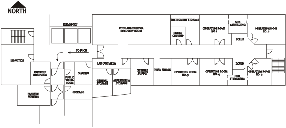 Diagram 3.1 Operating room suite
Anaesthetic nurse The anaesthetic nurse assisted the anaesthetists during high-risk operations. The nurse set up the lines, such as an arterial line or a central line, prepared the anaesthetic drugs, attached monitors to the patient and took samples of blood for the measurement of blood gases. Once the child was on bypass, the anaesthetic nurse might leave the OR until the patient was ready to come off bypass. On return to the OR, the nurse would co-ordinate bringing blood products into the room, preparing special drugs for administration and assisting with resuscitation as necessary. She would prepare the transfer sheet that accompanied the child from the OR to the Intensive Care Unit, identifying any problems or issues that arose during the course of surgery. She would then help prepare the patient for transfer, accompany the patient to the ICU, help set up the patient in ICU and give a report from the transfer sheet in order to make the transfer smoother. Perfusionists The perfusionists operated the cardiopulmonary bypass machine and administered cardioplegia (a treatment that stopped the heart during the actual operation). There were two perfusionists in each operation under discussion during this period. Residents Since the HSC was a teaching hospital, at times there would be residents or medical students, who would help either the surgeon or the anaesthetist. Surgical residents might be allowed to perform part of the procedure under the supervision of the surgeon, but most usually helped the surgeon by suctioning blood from the operative field or holding lines or instruments out of the surgeon's way to give him a clearer view. The resident was present primarily to learn. Anaesthetic residents might be permitted to administer drugs or insert tubes or lines, as directed and supervised by the anaesthetist. Usually, they also recorded (or charted) everything that occurred during the procedure. Cardiologists On occasion the surgeon would call for the cardiologist to enter the OR to help with identification of heart anatomy and discuss any issues with the case. The cardiologist would not scrub in but would wear OR garb and would then stand with the anaesthetist at the head of the OR table.
The teamThese persons constituted the operating room team. Historically, the surgeon was seen as the captain of the surgical ship, particularly since the patient, in medical terms, belonged to the surgeon, although that view has changed over recent years. As a result, the lines of command in the operating room also changed. The surgeon and the anaesthetist each were expected to have mastered separate medical specialties with distinct skills. Thus, neither the anaesthetist nor the surgeon was solely in charge. The surgeon was not responsible for matters relating to anaesthesia, while the anaesthetists were not responsible for the operation. They had a joint, if independent, responsibility for the patient's health and well-being. Both might discharge their duty by delegating certain aspects of the treatment to assistants who were properly qualified and experienced. However, it would be a mistake to simply focus on these two doctors. An operating room team depends on the skills, dedication and talents of all of its members. A team is more than a collection of highly skilled individuals, charged with working together. While it is essential for each team member to have the skills needed to accomplish the task at hand, a team needs a leader who is skilled in problem-solving and decision-making. As a whole, the team must be able to communicate effectively and resolve conflicts. This requires effective interpersonal skills. In a strong OR team, leadership is shared, problems are solved through discussion, members are accountable to each other and performance is measured collectively and regularly.
ChartsA medical chart is a record of what was done to a patient in the hospital. It is the record of medical proceedings and is an important-and, in most cases the only-source of information available when a patient dies, for those who then attempt to assess what happened. The office of the Chief Medical Examiner for the Province of Manitoba must by law and by policy be called whenever a child dies during or after a surgical procedure. In determining whether or not to direct that an autopsy be ordered or that an Inquest be called, one of the first steps a medical investigator takes is to look at the medical chart for information about the patient's treatment. If the information on the chart is not accurate or lacks a material fact, the Chief Medical Examiner's office is unable to come to a proper conclusion. The staff anaesthetist had the ultimate responsibility for charting on the anaesthetic record, but in some cases, a resident might do the charting. The anaesthetist charted the monitors used (such as types of lines), type of anaesthetic used, the drugs given, the dose and times they were given, the vital signs, the timing of bypass, intravenous fluids and blood products given and any problems that may have arisen. The perfusionists entered their information on the Perfusion Record. They recorded pump times and rates, line pressures, blood chemistry and blood gas values, information on clotting time, the delivery of heparin, cardioplegia doses and any blood products given. The circulating nurse was responsible for recording on the OR Count Sheet the OR times, the staff involved in surgery, the procedure performed and which specimens were sent for testing. A vital part of the record was the outcome of the needle, sponge and instrument count. To determine this, a count was made by the scrub nurse and the circulating nurse together. The surgeon did not do any record keeping during surgery. However, at the end of the operation, the surgeon dictated an operative report. A copy was placed in the patient's hospital record. There did not appear to be any systematic evaluation of surgical outcomes. Although the duration of each case and other factors were noted at the time, there was no process of tracking common indicators. These indicators could include duration of use of the heart-lung machine, duration of total circulatory arrest, volume of blood loss, number of units of blood transfused and number of blood components transfused: e.g., red blood cells, plasma and platelets.
The three phases of the operationEvery cardiac operation had three phases: pre-surgical, surgical and post-surgical. Sometimes these were quite distinct. At other times, because of the urgency of the operation and the condition of the patient, the first two phases might appear to occur almost simultaneously. The pre-surgical phase The pre-surgical phase started with the arrival of the anaesthetist. After suitable preparation, including attaching monitors, the patient would be anaesthetized, then positioned on the table for the operation. Special padding would be placed to protect the child. The area of the operation (also known as the 'surgical field') would be washed (or 'prepped') and then draped with sterile towels, ready for the first incision. These procedures were undertaken in the operating room. The anaesthetist largely performed the first step, preparing and anaesthetizing the child. The scrub nurse, often assisted by the surgeon, would do the prepping and draping of the patient. While these steps took place roughly in the order described, certain portions of these steps might be undertaken simultaneously or were completed in stages throughout the process of the start of the anaesthetic. Arrival of the anaesthetist The anaesthetist arrived in the operating room at least 30 minutes before the patient was scheduled to arrive. In high-risk cases the anaesthetic nurse and a respiratory technologist would assist the anaesthetist. The technologist would check equipment, such as monitors, while the nurse, under the direction of the anaesthetist, helped prepare necessary drugs and intravenous (IV) lines. The patient and parents After the arrival of the anaesthetist, the parents and child were brought to the waiting room, which was outside the operating room. The patient might or might not have been given a sedative before going to the operating room. In the case of infants and small children, the parents would carry their child in their arms to the operating room door and deliver the child into the arms of the waiting OR nurse. Anaesthetic monitoring Monitoring was an essential part of anaesthesia for pediatric cardiac surgery. Before the induction of anaesthesia, the anaesthetist would usually set up monitoring equipment. This equipment included devices to monitor:
Oxygen saturation was also monitored. Oxygen saturation is a term that describes how much oxygen is in the blood. A normal saturation is 97-100 per cent. When air enters the lungs, oxygen moves from the air, into the lung tissues, through the walls of the very small blood vessels (capillaries) and into the blood. Some of the oxygen stays in the liquid (or plasma) of the blood. Most of the oxygen enters the red blood cells, where it combines with a special chemical, known as hemoglobin. The amount of oxygen that actually binds with the hemoglobin is known as the oxygen content. The amount of oxygen that could bind with the hemoglobin is known as the oxygen capacity. The ratio between the content (actual) and the capacity (possible) is the oxygen saturation. Oxygen saturation can be easily measured by using a special probe, called a pulse oximeter. The probe is simply clipped onto a finger (or toe or earlobe), just like clipping on a clothes peg. No needles are inserted into the patient. Instead, the probe uses a special light beam to measure the oxygen saturation. The pulse rate is also measured, as the device notes each time a pulse of blood is pumped into the finger (or toe), each time the heart beats. Most pulse oximeters make a beeping noise every time a pulse is detected. This beeping will increase or decrease in rate with the pulse. The pitch (or musical tone) of the beep will also change as the saturation decreases. Other monitors were:
In addition, extra monitoring would be provided by special lines inserted into other blood vessels, such as the jugular vein in the neck, in the form of a central line (see below). Not all the monitors would be attached or inserted until after the start of the anaesthetic. Some children would be well enough that only a few monitors were needed for induction. Other children might be too agitated to have all monitors attached or inserted until they were either well sedated or actually anaesthetized. In addition, other monitors, such as the urinary catheter (usually referred to as a Foley catheter), generally were not inserted until after the child was anaesthetized. The circulating nurse would insert the catheter to measure and collect urine output. The placement of lines There were at least two intravenous lines (or catheters) inserted into veins in either the child's hands or feet. These were referred to as peripheral IV lines because they were inserted into veins in the patient's extremities (or periphery). If the patient was to undergo intravenous induction of anaesthesia, a line would be inserted (or placed) to allow the delivery of the anaesthetic drugs. Depending on the nature of the case, other lines might be inserted for monitoring purposes. These could include an intra-arterial line for the continuous measurement of blood pressure and the taking of samples of arterial blood during the course of the operation. Common arterial catheter sites included the vessels of the wrist, the foot, the groin and, in newborns, the umbilical vein. Another line might be used to measure central venous pressure. (Central venous pressure reflects the filling pressure on the right side of the heart and blood volume status). In addition, central venous lines were often inserted to allow the rapid administration of drugs, fluids or blood and permit the insertion of pulmonary artery or pacing catheters. Sites used for placement of central venous catheters included the internal or external jugular veins, the femoral vein (in the groin), a vein in the elbow and the umbilical artery. Central venous catheters might also be placed directly into the right atrium during cardiac surgery (and were often termed 'RA lines'). In most cases, these lines were inserted by putting a needle through the skin and then threading the catheter through the needle. However, the lines could also be inserted by performing a 'cut-down', a procedure in which the skin was cut open and the vein or artery exposed, to make it easier to insert the catheter. The catheters were then connected to tubing that was connected to transducers to facilitate the reading of pressures. (A transducer is a small device that changes the mechanical energy from the monitored pressure to an electrical signal captured on the monitoring screen.) The anaesthetists generally used two transducers (one arterial and one venous). The surgeon might require three transducers: one for a right atrial line, one for a left atrial line (an LA line) and sometimes one for a pulmonary artery line (a PA line). Depending on the operation, these lines might be put in intra-operatively and/or post-operatively. (Intra-operative is the term that refers to events that occur during the operation.) As long as these lines were in place, the transducers were left attached and were used in the ICU. Induction and intubation The induction phase of anaesthesia was the process of rendering a patient unconscious by giving the patient intravenous drugs through an intravenous line or inhaled drugs through a mask attached to a tube from the anaesthetic machine. After the anaesthetic drugs had taken effect, the anaesthetist then inserted a special breathing tube down into the patient's windpipe (in medical terms, the trachea). The anaesthetist would not start the anaesthetic until receiving confirmation that the surgeon was in the hospital. The process of induction and intubation could then take from twenty minutes to two hours, depending on the condition of the child and any difficulties encountered. Induction The anaesthetist would have determined which drugs were to be used to anaesthetize the patient. Initially the anaesthetic would be administered either intravenously or as a gas that the child breathed in. (In medical terms, this special gas is known as an 'inhalational anaesthetic agent' because the drug is inhaled or breathed in.) Once the child had lost consciousness, the anaesthetic would be continued with both intravenous and inhaled drugs. These drugs included sedatives (such as midzolam), narcotics (such as fentanyl, sufentanil, alfentanil and morphine) and an inhalational agent (such as halothane or isoflurane). In addition, the child might also breathe in some nitrous oxide (known to many people as 'laughing gas'). The state of anaesthesia was maintained with further intravenous drugs and inhaled gases. For these cardiac operations, the patients were also given specific anaesthetic drugs to weaken or relax their muscles. This helped the anaesthetist to be able to place the breathing tube into the trachea and then allowed the surgeon to operate on the chest without tearing the normally tight muscles of the chest. These special drugs were known as muscle relaxants. They included pancuronium, vecuronium and atracurium. Intubation Intubation refers to the placing of one tube inside another. The intubation that was referred to in a pediatric cardiac case was called endotracheal intubation. In this process a tube was inserted into the patient's trachea or windpipe, through either the nose or the mouth. The tube guaranteed a passage or 'airway' through which the patient could be ventilated. The tube also helped to prevent unwanted substances from going into the patient's lungs. To make intubation easier, the child was given a muscle relaxant. As a result, the child would not be able to breathe and therefore required artificial ventilation. (This ventilation delivered oxygen to the lungs and removed carbon dioxide, in the same way that normal breathing did, except that the breathing was done for the patient.) First, the anaesthetist would hold a rubber mask over the patient's face, making an airtight seal. Then the anaesthetist would squeeze a special rubber bag that was attached by a rubber tube or hose to the mask. When this bag was squeezed, oxygen would be pushed from the bag, through the tube and mask, into the patient's nose and mouth and down into the lungs. Once the muscle relaxant was fully effective, the anaesthetist would insert the breathing tube in through the patient's nose or mouth and down into the windpipe or trachea. (In medical terms, this procedure is known as 'performing tracheal intubation'.) Once this tube was positioned and secured (usually with some tape), the anaesthetist would use a machine called a ventilator to breathe for, or artificially ventilate, the patient's lungs. This ventilation was delivered through the breathing (or endotracheal) tube. Positioning, padding, prepping and draping the patient The positioning, padding, prepping and draping of a patient could take half an hour. Positioning Most operations were carried out with patients lying on their backs. Occasionally, some operations were performed with the patient lying on his or her side. This meant that the patient had to be carefully turned and positioned, after the start of the anaesthetic and the placement of all the lines and monitors. Padding The patient's head and face and the lines would be padded for protection during surgery. Padding was not a sterile procedure and was done by the circulating nurse, anaesthetic nurse and anaesthetist. In some cases, the anaesthetist would pack ice around the patient's head and neck to cool the brain. This was done to protect the brain when there was to be a complete stoppage of both the heart and the heart-lung machine. Prepping For heart surgery, the site of the operation (or 'operative field'), which was the chest, was then washed with a special antiseptic solution. The surgeon, or his assistant or the scrub nurse, would do this. Draping Because draping was a sterile procedure, it was done by the surgeon (or his assistant) and the scrub nurses. They placed sterile cloths or drapes over all but the operative field. A small, flexible screen would separate the patient's head from the surgical field. This screen would be draped with a sterile cloth to denote the barrier between the sterile surgical field and the patient's head, to which the anaesthetist would usually have access. This sterile drape was hung or draped in a manner to maintain the sterility of the area where the operation would be carried out. The surgical phase The surgical phase would start with the first incision and proceed until the completion of the operation. Pediatric cardiac operations were either closed or open procedures. Closed-heart surgery Closed-heart surgery was that surgery in and around the heart that did not involve having to stop blood flow to the heart. It therefore did not involve having to bypass blood around the heart or involve having to stop the heart from beating. Generally, closed-heart procedures were less risky than open procedures because the process of bypassing the heart did not need to be undertaken. Nonetheless these procedures did involve their own risks and some were as risky as or riskier than some of the simple open-heart procedures. Open-heart operations Open-heart surgery required the use of a cardiopulmonary bypass (CPB) machine. The CPB machine was a pumping device that diverted blood away from the heart, and returned it to the body at a point beyond the heart. Because the heart itself is essentially a pump, when it was stopped or blood was diverted around it, the CPB machine had to take over the pumping function of the heart. The CPB machine also allowed the blood that was pumped to pick up oxygen and be cleansed of carbon dioxide. These two essential functions were made possible through a membrane oxygenator that allowed oxygen to move into the blood and allowed carbon dioxide to move out of the blood. The use of CPB was an important development in cardiac surgery. It has allowed surgeons to operate on the heart and major blood vessels while the patient's systems were otherwise maintained. However, a patient could not be maintained on CPB indefinitely. A lengthy bypass would affect many aspects of a patient's system. The lungs would become wetter and the small airways would collapse. The liver and kidneys could be damaged. The heart could accumulate water and become swollen (or in medical terms 'edematous'). As a result, the heart could become less and less compliant and the heart muscle itself could also be damaged, contracting poorly, because of difficulties with techniques intended to protect the heart. (In medical terms, these techniques were known as 'cardiac preservation'.) The patient's blood could also be damaged. Increased CPB times could also increase the risk of air in the cannula lines, creating an airlock in the patient's blood vessels. Open procedures could also involve stopping the heart from beating for a while. This was referred to as total circulatory arrest (or TCA). While open procedures were considered the riskiest of the cardiac procedures, those requiring TCA placed the patient at greatest risk. When the beating of the heart was stopped, the CPB machine was also stopped and blood flow through the body was stopped. In a procedure known as deep or profound hypothermia arrest (DHA or PHA) the body was invariably cooled to as low a temperature as possible (usually around 16-20 degrees Celsius) and the head was packed in ice. Despite such measures, however, TCA was a time-limited procedure. Most medical experts agreed that a period of 30 minutes of TCA would not likely impair brain function and that 45-60 minutes was the safest maximum period of TCA allowed. Beyond that time, most experts agreed that the chances of brain damage or neurological impairment increased dramatically. There were three stages to an open-heart pediatric cardiac operation. These were pre-bypass, on-bypass and post-bypass. Pre-bypass The pre-bypass stage started when the surgeon made the initial incision and then proceeded to open the patient's chest. Before doing this, the surgeon made it clear that he was ready to start and gained assurances that all other members of the team were ready. The patient's body might react to the initial incision in ways that could not always be predicted. The anaesthetist then needed to respond to these changes by increasing or decreasing the amount of anaesthetic or administering other drugs or fluids, depending on the patient's reaction. For this reason, it was essential that the rest of the team be alerted to and prepared for the surgeon's activities. There were certain procedures the surgeon needed to perform before going on-bypass. These could be addressing palliative repairs that had been done previously, carrying out repairs that could be done without going on-bypass (thus shortening the length of the bypass) and finally inserting the cannulas into the heart (cannulation) and attaching them to tubing for connection to the CPB machine. The major elements of the pre-bypass portion of the surgery were:
Repairs In either open or closed-heart surgery, it was at this point in the operation that the surgeon attempted to carry out some of the necessary repairs to the heart. (These repairs were briefly outlined in Chapter 2.) It should be again emphasized that these repairs were extremely difficult to execute, given that the surgeon was working on a very small, very variable structure that might be moving with every heartbeat and every breath if CPB was not being used.
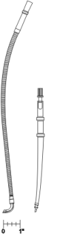
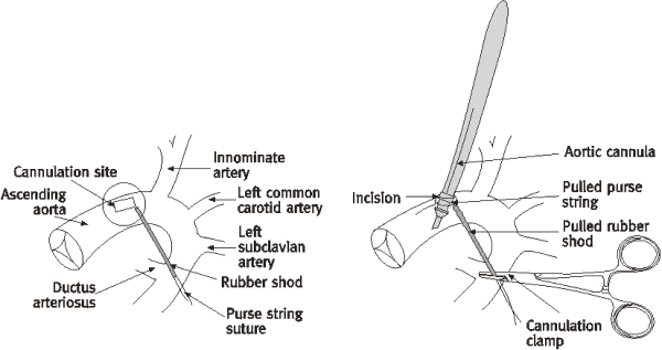
Heparinization In order to go on bypass, the patient's blood had to be treated with a drug called heparin to prevent it from clotting. If blood was passed through the heart-lung machine without heparinization, it would begin to clot immediately. This would be fatal to the patient. Before administering heparin, the perfusionists measured the child's activated clotting time (ACT). After the heparin was given, a second ACT measurement was taken to ensure that the patient was fully heparinized. After this, the clotting time was measured at regular intervals during bypass to ensure that it stayed above a set limit. The patient could then be cannulated, cardioplegia could be given and CPB could be established. Cannulation The pediatric cannulas were tiny catheters used to connect the patient's heart to lines (or tubes) that were connected to the cardiopulmonary bypass machine (CPB). The cannulation procedure required the surgeon to insert the cannula into the major blood vessels (the inferior and superior vena cavas and the aorta) on either side of the heart. The cannula that was inserted into the aorta was referred to as the aortic cannula, while the cannulas inserted into the inferior and superior vena cavas were referred to as the venous cannulas. The venous cannula and the aortic cannula differed in shape to accommodate differences in the structure of the blood vessels into which they were inserted. It was important to carefully place the cannulas and the lines running to the CPB machine to ensure that they did not become dislodged. The aortic cannula, which connected the CPB machine via tubing to the aorta, was placed first, in case the patient needed to be transfused quickly. It was through this line that oxygen-rich blood was pumped. It was often the practice to place two cannulas in the vena cava, one in the SVC and one in the IVC. Oxygen-poor blood flowing from the body was then directed away from the heart, through these cannulas and to the CPB machine. The actual process of inserting the cannula involved the following steps:
At this point the cannula would be connected to the line from the CPB machine.
Cardioplegia It was extremely difficult, if not impossible, to operate safely and quickly on a beating heart, particularly a tiny heart that might be beating at more than 100 times a minute. The heart therefore had to be stopped from beating. This was accomplished by the infusion of a high-potassium solution into the heart's coronary arteries, from a machine known as a blood cardioplegia device. The stopped heart was deprived of oxygen and nutrient-rich blood and was at risk of damage. Injecting cooled oxygenated blood (that was mixed with the potassium solution) into the coronary arteries therefore provided the stopped heart with protection from this damage. The solution injected was known as a cardioplegic solution and provided what was known as myocardial protection. Because the cardioplegic mixture was extremely cold (approximately six degrees Celsius) it reduced the heart's metabolic state to the very lowest possible point once it had been arrested. Cardioplegia allowed the heart to maintain its energy stores and could also prevent the heart from beating for twenty to thirty minutes before an additional dose was required. During bypass, the EKG monitor would normally show a flat line. Any sign of electrical activity represented the need for further cardioplegia. It might be necessary to provide an additional dose of low potassium solution to maintain the arrest. To assist in this it was the perfusionist's role to remind the surgeon at intervals of approximately 20 minutes when the last cardioplegia had been provided. The surgeon decided, in consultation with the perfusionist, if another dose was to be given. The perfusionist would actually switch on the infusion, administer the solution and record the volume given. Acidosis One of the conditions that often occur in children with congenital heart problems is acidosis. This is the accumulation of an abnormally high level of acidity, or low level of alkalinity, in the body fluids, including the blood. There are two general types of acidosis: metabolic acidosis and respiratory acidosis. Metabolic acidosis occurs when the body takes in too much acid, makes too much acid, or does not excrete enough acid through the kidneys. Respiratory acidosis occurs when the body retains carbon dioxide, which the body senses as an acid (carbonic). Acid can come from increased production, usually of lactic acid, or when the body shifts into 'anaerobic' metabolism. This most often occurs when blood flow to the tissues decreases, and the tissues then shift from aerobic (with oxygen) metabolism to anaerobic (without oxygen) metabolism. When tissues are poorly perfused with blood, they do not use as much lactic acid as they normally do; this also contributes to an increase in the amount of lactic acid in the blood. In addition, severe acidosis may itself lead to reduced blood flow to the tissues, because the acid impairs the heart's ability to contract normally. As the acidosis becomes worse, the patient starts to breathe faster and take deeper breaths, as the body tries to compensate for the excess acid. Finally, the heart weakens and the patient may progressively lose consciousness. There are a number of approaches to treating acidosis. These involve increasing blood flow to the tissues by strengthening the heart, countering the acidosis by treating the child with bicarbonate and providing general supportive treatment in the form of oxygen and fluids. On-bypass Following cannulation and the administration of cardioplegia, the bypass machine was connected to the patient through the cannulas and took over the heart's functions. This was the start of bypass and was known as 'going on pump'. After going on pump, the surgeon might need to operate inside the heart (referred to as intracardiac) and would therefore need to stop the heart. The surgeon would take a special instrument, called an aortic cross-clamp, and use it to pinch or seal off the aorta. No blood from the heart could therefore go past the clamp and out to any part of the body, including the brain. The blood supply to the body and brain was then entirely from the CPB machine via the aortic cannula, with blood returning to the CPB machine via the venous cannulas, without passing through the heart's ventricles. The length of time that the cross-clamp was completely applied to the aorta would be recorded. This time would be known as 'cross-clamp time'. Another term was 'total ischemic time', which meant the time that the body was without blood flow and oxygen delivery during periods of circulatory arrest and profound hypothermia. (In medical terms, lack of blood flow is known as 'ischemia'.) The heart would then be stopped and preserved with cardioplegic solution. The end of 'cross-clamp time' would be when the cross-clamp was partially or completely released. Even partial release of the clamp would allow some oxygen-containing blood to flow to the brain and other organs. Blood Throughout the bypass portion of the procedure, blood would accumulate in the surgical field. The surgeon or his assistant would suction this blood away to allow the surgeon to see the operative site. This blood was filtered, added to the reservoir in the CPB machine and returned to the body. When required, additional blood could be transfused into the patient, through either the CPB pump or the IV lines. When necessary, bags of packed red blood cells were brought into the OR. These bags would usually be kept in a special refrigerator down the hall from the operating room. Before any blood was administered to the patient, it would be double-checked by members of the OR team to ensure that it matched the patient and the patient's blood type. Some blood products, such as fresh frozen plasma, would have to be thawed if they were required, while others, such as blood platelets, would have to be kept warm and agitated gently, to prevent them from congealing. The HSC did not, as a matter of course, maintain a large supply of blood at the hospital. Blood products were kept by the Red Cross at its Winnipeg facility. Whenever a surgeon ordered blood products for an operation, the hospital had to request those products from the Red Cross ahead of time. The Red Cross, in turn, would approve the request and then arrange for delivery of the blood to the hospital in time for the procedure. If an emergency occurred and blood was needed quickly, the hospital did have a small supply of blood it could use. However, occasionally it was necessary to arrange for blood products to be delivered specially from the Red Cross-often by taxicab. Throughout the operation the patient's blood would be tested (usually hourly) to check the hemoglobin concentration. The times when the blood was drawn and when the tests were conducted were usually noted on the chart. Ventilation The anaesthetist was responsible for ensuring that a patient was properly ventilated during the operative procedure. Once the patient was on bypass, the anaesthetist would stop ventilating the patient, since the blood would receive its oxygen from the CPB machine. During these times, the ventilator would be turned off and the oxygen supply from the anaesthetic machine would be turned down, with just a small amount of oxygen being delivered down the endotracheal tube. With the ventilator turned off during surgery, the lungs would not balloon into the area around the heart and block the surgeon's view of the heart. Before weaning The process of discontinuing bypass and allowing the heart to take over the pumping of blood throughout the patient's system was referred to as 'weaning from bypass'. In order for the patient to be weaned from bypass successfully, everyone involved in the operation-the surgeon, anaesthetist, perfusionists and nurses-had to work in a co-ordinated fashion. Before weaning from bypass, the anaesthetist would suction the endotracheal tube, to remove any secretions that might have accumulated. In addition, a drug might be given to those patients whose airways might be sensitive to suctioning. (In medical terms they were said to have 'reactive airways'.) Monitoring lines After an operation was completed, it was vital for the doctors and nurses caring for the child to be able to monitor the state of performance of the child's heart. To facilitate this, the surgeon would place monitoring lines in the child's heart before closing the chest. These monitoring lines were referred to as 'transthoracic lines' and were eventually connected to monitors in the ICU. The surgeon would have to decide whether or not to place these monitoring lines before weaning the patient from bypass or if the lines could be placed after bypass was discontinued. Usually the surgeon would insert the transthoracic lines in the atria and the pulmonary artery after decannulation (removing the cannula). These lines measured atrial and pulmonary artery pressures. The scrub nurse would hand the surgeon a sterile set of high-pressure lines. After inserting one end of the line into the heart chamber, the surgeon would hand the other end of the line to the anaesthetist, who would connect it to a transducer. The line with the transducer attached would then be available for the ICU staff to use when the patient arrived in the unit. Weaning from bypass Before the patient was weaned from bypass, the surgical team had to ensure that the patient was in optimal condition. Close attention had to be paid to the monitors and the condition of the repair. The anaesthetist had to ensure that the patient was well anaesthetized and relaxed. The anaesthetist also had to be ready to deal with any consequences that might arise from coming off bypass. This included having special drugs ready to support the circulation. If there were concerns about the heart's ability to contract forcefully, inotropes (drugs that helped the heart to contract) might be given. If the patient's blood pressure was too low, the anaesthetist might administer a vasoconstricting drug, such as neosynephrine, to raise the pressure. If the blood pressure was too high, vasodilators (drugs that dilated blood vessels) would be used to dilate the vessels and thereby reduce the pressure that the heart was pumping against. Coming off bypass Patients were 'taken off' bypass in a gradual but sequential manner. The patient had to be rewarmed to normal temperature, the heart once more had to start beating, ventilation of the patient was restarted, the perfusion was brought to an end, pacing wires were inserted, the heparin was reversed and the heart cannulas were removed from the patient. Rewarming The rewarming period was begun by increasing the temperature of the blood being perfused through the arterial line to approximately 8-10 degrees Celsius above the venous blood temperature. Often vasodilators were given to produce a smooth and even rewarming process. Starting the heart Before taking the patient off bypass, the surgeon needed to start the patient's heart beating again at a rate that was appropriate. In some cases, the heart would not beat fast enough. In other cases, the heart might need to be stimulated (with a small electrical shock) in order to start beating. Restarting ventilation Ventilation was usually restarted during rewarming, once the heart was closed. Stopping perfusion When the cardiac action had reached a satisfactorily vigorous state, bypass would gradually be discontinued. Once the heart had taken over from the machine, the venous lines connecting the CPB to the heart were clamped. Insertion of pacemaker wires The surgeon would insert pacemaker wires at this point. The pacing wires were put in, even if the operation seemed to have gone well. Drugs might be administered to help increase the heart rate. Reversing the heparin With the patient still heparinized, the team would then wait and monitor blood pressure and other vital signs. At this point an echocardiogram might be done to assess the repair. If all signs were appropriate, protamine would be administered to reverse the heparin. When the protamine was given, the anaesthetist needed to advise the perfusionists, because the perfusionists had to stop suctioning blood from the patient's chest cavity. If they did not, blood containing protamine might then be taken into the pump, with the risk of a blood clot in the pump. Decannulation When the surgeon was satisfied with the movement of blood through the heart, with the pressures in the chambers of the heart, and the blood pressure and heart rate (medically referred to as hemodynamics), the perfusion cannulas could then be removed. The cannulas were still in place when the protamine was given. However, with the administration of protamine, the chance of clots forming around the cannulas increased and the cannulas had to be removed. The surgeon would first take out the venous cannulas, leaving the aortic cannula as the last to be removed. It was important that the surgeon inform the operating team when he was about to remove the cannulas, as this could cause hemodynamic changes. In addition, there could be bleeding and the patient might need to be transfused quickly through the aortic cannula. Post-bypass Once the cannulas were removed, the patient was then completely separated from the bypass machine and was considered as being 'off pump'. The surgeon would then check the suture lines for any bleeding and ensure that any shunts or other tubes were patent. If all was satisfactory, he could then begin to close the patient's chest. A chest X-ray might be taken at this time to give the surgeon and anaesthetist information about the condition of the lungs and the placement of tubes and lines. At the same time, the nursing staff would complete their charting and phone the intensive care unit to inform them as to the expected time of arrival of the patient and the patient's general condition. The post-surgical phase In the post-surgical phase, the patient would be prepared for transport to the intensive care unit. Once the anaesthetist and surgeon had determined that the child was stable enough to transfer, the patient would be moved to the unit and cared for there.
|
| Current | Home - Table of Contents - Chapter 3 - Special Issues in Pediatric Medicine |
| Next | Post-surgical care |
| Previous | Events leading up to the day of surgery |
| Section 1 | Chapter 1 - Introduction to the Issues |
| Chapter 2 - Pediatric Cardiac Issues | |
| Chapter 3 - The Diagnosis of Pediatric Heart Defects and their Surgical Treatment | |
| Chapter 4 - The Health Sciences Centre | |
| Section 2 | Chapter 5 - Pediatric Cardiac Surgery in Winnipeg 1950-1993 |
| Chapter 6 - The Restart of Pediatric Cardiac Surgery in 1994 January 1, 1994 to May 17, 1994 |
|
| Chapter 7 - The Slowdown May 17 to September 1994 |
|
| Chapter 8 - Events Leading to the Suspension of the Program September 7, 1994 to December 23, 1994 |
|
| Chapter 9 - 1995 - The Aftermath of the Shutdown January to March, 1995 |
|
| Section 3 | Chapter 10 - Findings and Recommendations |
| Appendix 1 - Glossary of terms used in this report | |
| Appendix 2 - Parties to the Proceedings and counsel | |
| Appendix 3 - List of witnesses and dates of testimony | |
| Diagrams | |
| Tables | |
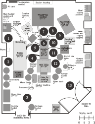 Diagram 3-2