|
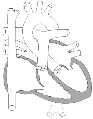
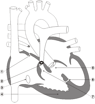
The decision to operateGiddins saw Alyssa at the VCHC on March 18, 1994. An echocardiogram performed that day revealed Tetralogy of Fallot, with:
The diameter of the bicuspid pulmonary valve was eight millimetres. The bilateral proximal pulmonary arteries were narrowed to five to six millimetres in diameter.
A chest X-ray done on March 18 showed that Alyssa had an enlarged heart. According to radiologist Dr. A.C. Patton, there was no evidence of acute pulmonary infiltrates. This is the term for the white areas on X-rays of the lungs. Infiltrates are also referred to as densities. The absence of infiltrates indicated that the lungs were normal. In subsequent testimony, Dr. Martin Reed, the head of pediatric radiology at HSC, indicated that he believed the X-ray actually revealed a mild abnormality. Giddins advised that surgery was required in the near future. While the Stills had been told that an operation would be necessary, they did not expect that it would take place so quickly. Donna Still testified that the nature of the problem was explained. She was left with the sense that she had no real choice but to proceed with the operation. While Odim was not present at this meeting, Still testified that Giddins told her Odim was one of the best surgeons in North America. She said that she was left with the impression that, while Odim was new to the hospital, he had worked-as opposed to trained-in Boston and Montreal. She also said that Giddins indicated to her that the success rate for this procedure was 95 per cent. Four days after Giddins met with the Stills, Alyssa's case was presented at a CVT conference involving Odim and Giddins, as well as staff from the Heart Centre. After the conference, Odim outlined his conclusions in a March 22 letter to Giddins. Odim described Alyssa as a four-month-old child whose evaluation confirmed the picture of Tetralogy of Fallot. She had no obvious symptoms of heart problems and was not taking any medications. He mentioned that the chest X-ray showed clear lungs and a slightly enlarged heart. His conclusions differed from Giddins's. Odim had detected an atrial septal defect, while Giddins reported that there was an intact atrial septum. Odim wrote that his view was that the child needed early repair and that he would be happy to discuss the operative approach and risks with the family (Exhibit 11, page STI 1). On March 22 Giddins wrote to Derbyshire that "Alyssa has Tetralogy of Fallot, with an atrial septal defect and significant obstruction at both the sub-pulmonary and pulmonary valve level." (Exhibit 11, page STI 2) Giddins stated that "Her muscular ventricular septal defect is likely not significant, and overall my impression is one of anatomy highly favourable for definitive early correction. The nature of the problem was explained to the family, as was our tendency to operate before six months of age, a concept they agreed with." (Exhibit 11, page STI 2)
|
| Current | Home - Table of Contents - Chapter 6 - The decision to operate |
| Next | Consent |
| Previous | Background and diagnosis |
| Section 1 | Chapter 1 - Introduction to the Issues |
| Chapter 2 - Pediatric Cardiac Issues | |
| Chapter 3 - The Diagnosis of Pediatric Heart Defects and their Surgical Treatment | |
| Chapter 4 - The Health Sciences Centre | |
| Section 2 | Chapter 5 - Pediatric Cardiac Surgery in Winnipeg 1950-1993 |
| Chapter 6 - The Restart of Pediatric Cardiac Surgery in 1994 January 1, 1994 to May 17, 1994 |
|
| Chapter 7 - The Slowdown May 17 to September 1994 |
|
| Chapter 8 - Events Leading to the Suspension of the Program September 7, 1994 to December 23, 1994 |
|
| Chapter 9 - 1995 - The Aftermath of the Shutdown January to March, 1995 |
|
| Section 3 | Chapter 10 - Findings and Recommendations |
| Appendix 1 - Glossary of terms used in this report | |
| Appendix 2 - Parties to the Proceedings and counsel | |
| Appendix 3 - List of witnesses and dates of testimony | |
| Diagrams | |
| Tables | |