|
Autopsy findingsThe final report of the autopsy, which was conducted by Dr. Susan Phillips of the HSC, was released on June 29, 1994. The autopsy did not identify a cause of death. Her report indicated that the repair was intact. In addition she identified:
Various consulting witnesses before this Inquest discussed these findings, which bear comment. It should be noted that these witnesses not only reviewed Phillips' report; they also examined Alyssa's heart.
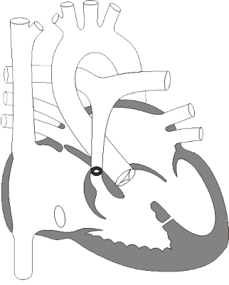
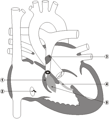
The presence of a suture in the coronary sinus In his operative report, Odim stated that he closed a small VSD with an interrupted suture. In her autopsy report, Phillips stated that she could not identify the VSD or the suture that Odim reported placing over it. This is disturbing. It is very unlikely that Phillips would not have been able to detect such a suture if it was indeed present in the heart. At the same time, Phillips did record finding an interrupted suture "in the region of the coronary sinus. The coronary sinus is patent." (Exhibit 11, page STI 24) Odim recorded making use of an interrupted suture in closing the VSD. This gives rise to the possibility that the suture found in the region of the coronary sinus was in fact the suture that Odim intended to place over the VSD. While this appears possible, there is a larger question. Did this suture close the sinus? The coronary sinus drains blood from the coronary veins, which in turn drains the heart muscle. If the coronary sinus were sutured closed, then the heart muscle would not drain properly. Taylor testified that the effect of a closed coronary sinus depends on the extent of the connection between the venous system and the ventricles of the heart. If there were few or no direct connections, an obstruction of the coronary sinus would interfere with venous return from the heart muscle. This could cause the heart to swell, leaving the child with edema or congestion in the heart muscle. The outcome would be impaired function of the heart. Because the connections are microscopic, it was not possible in Alyssa's case to determine whether there was adequate accessory venous drainage into the ventricles or the atria itself (Evidence, pages 43,059-43,060). In her report, Phillips stated that the sinus was patent or open. Phillips also indicated that she rechecked for patency before testifying for the Inquest and again found it patent. She checked for patency by inserting a small metal probe into the opening-the sinus was described as probe patent. In his initial report for the Inquest, Taylor concluded that the suture did not close the sinus. However, when Taylor testified, he offered a different opinion: Of note here is a pledget suture which is placed near the coronary sinus, which is right here. And in my report I just said that it was near by, but looking at my notes from the time that I did the examination, I noted that it actually closes the coronary sinus which is right here. (Evidence, page 43,059) Taylor also testified that, in his opinion, the closed coronary sinus was not a significant adverse factor but rather a minor factor (Evidence, page 43,075). Cornel, however, was concerned by the presence of the suture at the orifice of the coronary sinus, because it might have contributed to the failure to resuscitate following cardiac arrest (Exhibit 353, page 39B).
Necrosis is the death of an area of tissue and may follow a wide variety of injuries. The injury activates enzymes in the cells that destroy the damaged cells, resulting in dead or necrotic tissues. There is usually a lapse of time between the injury and the development of necrosis. Pathologists can often detect the signs that indicate this lapse of time. Phillips recorded that microscopic examination of the heart revealed patchy subendocardial contraction band necrosis of the myocardium of both ventricles. Alyssa therefore had necrotic or dead areas of both ventricles of her heart (or myocardium). This necrosis specifically affected the subendocardial region of the heart muscle-the inner third to half of the ventricle. In addition, she had necrosis in the form of contraction bands. These microscopic structures in the heart muscle appear like bands of dead tissue and are formed by over-contraction of the muscle cells. The presence of any necrosis decreases the heart's ability to conduct electricity normally and to pump normally. According to Phillips's testimony, there is normally a lapse of about four hours between the time of the injury and the development of contraction band necrosis. Taylor testified that the type of necrosis that Alyssa experienced usually indicates inadequate myocardial protection during open-heart surgery, although it can also occur with cardiac arrest and with the administration of certain drugs, such as adrenalin. He said: In this setting the thing I would be thinking of as far as trying to put everything together would be some injury suffered as a result of unsatisfactory or insufficient myocardial protection during the long bypass procedure. (Evidence, pages 43,080-43,081) In Taylor's opinion, the necrosis developed during surgery and was a sign that the heart had not been provided with sufficient myocardial protection. Taylor also testified that the fact that Alyssa had prolonged surgery, needed inotropic support and was acidotic were all indications that acute myocardial impairment had occurred during surgery.
The autopsy revealed widespread retention of fluid, or edema. This may be an indication that the heart's capacity to pump had been compromised. The autopsy detected:
Subcutaneous edema Subcutaneous edema was particularly apparent in Alyssa's face and eyelids. This form of edema does not necessarily signify that there has been excessive damage to the child during surgery. Rather, the presence of edema here only signifies that the patient has retained a considerable amount of fluid. Pulmonary edema Microscopic examination of lung tissue revealed diffuse areas of congestion and patchy areas of edema fluid in the air sacs (alveoli). Pulmonary congestion occurs when the blood vessels of the lung are so filled with blood that they are distended. Taylor testified that: Edema fluid in the lungs indicates that there is some left heart failure or injury to the lining of the small blood vessels in the lungs, such that blood cells can't leak out but fluid from the lung can leak out. (Evidence, page 43,078) Taylor wrote in his report that pulmonary congestion and edema were significant adverse factors in this case (Exhibit 336, page 5.1). Pulmonary congestion and edema can develop very quickly and might have been as a result of Alyssa's cardiac arrest, rather than the cause of it. Cerebral edema There was also swelling or edema of the brain. Microscopic examination of the brain revealed evidence of acute hypoxic/ischemic injury or new brain damage as a result of a lack of oxygen and blood flow. Odim and Taylor differed as to the source of the damage. Odim was not surprised that there was brain damage, given the resuscitative measures undertaken. He testified that: It's not unusual after cardiac arrest to have lack of cerebral blood flow because the heart isn't pumping for 40 minutes. (Evidence, page 24,929) Taylor believed that the brain damage occurred most likely during the operation: There is insufficient time for the changes to occur from when she arrested and died to when the autopsy was done. It had to be 12 hours or even longer before that. (Evidence, page 43,095) The reduction in blood flow through the coronary sinus and the presence of contraction band necrosis in the heart meant that less oxygen could be delivered to the brain, leading to brain damage. The weight of the evidence suggests that both the heart and brain damage did occur during surgery. Myocardial edema Taylor also testified that he found mild myocardial edema. This can occur when fluid collects between the cells of the heart muscle. Taylor testified: I consider that a significant factor because I believe that myocardial edema in a heart that has been subjected to open heart surgery and bypass can occasionally cause problems. And I make that statement on the basis of somewhat similar cases to Alyssa that I encountered in the past, where really the only finding was myocardial edema. I think myocardial edema occurs to greater or lesser degree after heart surgery almost always. But sometimes, depending on the metabolic state of the child, other anatomical factors, the myocardial edema may cause the heart to become dysfunctional and put it at jeopardy for something like an arrhythmia. (Evidence, page 43,077) Hudson did not think that myocardial damage or dysfunction caused the cardiovascular collapse: Poor myocardial function would likely have been evident sooner, and would be more likely to be manifest as progressive deterioration rather than a sudden, catastrophic event. Therefore, I do not feel that poor myocardial function was the primary cause of this child's death. (Exhibit 307, page 5.19) In response to questioning about the widespread edema, Odim indicated it was a result of a variety of factors: including bypass and medication. It was not, he felt, unusual, given the circumstances (Evidence, pages 24,928-24,931). While there is truth to this, as several witnesses pointed out, events during surgery may have unnecessarily contributed to the development of this edema. The repair to the ventricular septal defect In his examination of Alyssa's heart for this Inquest, Taylor identified a two-millimetre leak in the Dacron patch closing the ventricular septal defect. In his testimony, he said that it was not possible to determine if the leak was caused before or after Alyssa's death.
|
| Current | Home - Table of Contents - Chapter 6 - Autopsy findings |
| Next | Cause of death |
| Previous | Post-operative course |
| Section 1 | Chapter 1 - Introduction to the Issues |
| Chapter 2 - Pediatric Cardiac Issues | |
| Chapter 3 - The Diagnosis of Pediatric Heart Defects and their Surgical Treatment | |
| Chapter 4 - The Health Sciences Centre | |
| Section 2 | Chapter 5 - Pediatric Cardiac Surgery in Winnipeg 1950-1993 |
| Chapter 6 - The Restart of Pediatric Cardiac Surgery in 1994 January 1, 1994 to May 17, 1994 |
|
| Chapter 7 - The Slowdown May 17 to September 1994 |
|
| Chapter 8 - Events Leading to the Suspension of the Program September 7, 1994 to December 23, 1994 |
|
| Chapter 9 - 1995 - The Aftermath of the Shutdown January to March, 1995 |
|
| Section 3 | Chapter 10 - Findings and Recommendations |
| Appendix 1 - Glossary of terms used in this report | |
| Appendix 2 - Parties to the Proceedings and counsel | |
| Appendix 3 - List of witnesses and dates of testimony | |
| Diagrams | |
| Tables | |