|
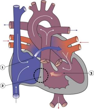
Tricuspid atresiaAtresia means the absence or closure of a normal body opening or tubular structure. In tricuspid atresia, the tricuspid valve is missing, preventing blood from flowing from the right atrium into the right ventricle. Because the right ventricle has no blood to pump, it remains small and underdeveloped. In this condition, the child's survival depends on the presence of two septal defects: an atrial septal defect (ASD) and a ventricular septal defect (VSD). The ASD allows the venous blood to flow from the right atrium into the left atrium. There, venous blood mixes with oxygen-rich blood from the lungs, flows to the left ventricle, into the aorta and out to the body. The rest of the mixture is pumped from the left ventricle through the VSD into the right ventricle, and on through the pulmonary artery back to the lungs. Sometimes the baby will also have other cardiac defects. These include transposition of the great arteries (to be discussed later), coarctation of the aorta or pulmonary atresia. The majority of babies with tricuspid atresia are cyanotic at birth. These children will require surgery to place a tube (known in medical terms as a shunt), which will provide an increase in blood flow to the lungs. Other children have too much blood flowing to the lungs and need a procedure called pulmonary artery banding. In this procedure, the surgeon places a band around the pulmonary artery to narrow it, and to reduce the blood flow and resultant high pressure in the lungs. In other children the surgeon connects the right atrium and the pulmonary artery and closes the ASD. This procedure is known as a Fontan, named after the doctor who first perfected the procedure. The Fontan repair allows blood to flow passively from the right atrium to the pulmonary circulation without being pumped by the right ventricle. The functioning of this repair relies totally on the preservation of a low resistance in the pulmonary arterial vessels.
|
| Current | Home - Table of Contents - Chapter 2 - Tricuspid atresia |
| Next | Pulmonary atresia |
| Previous | Pulmonary stenosis and aortic stenosis |
| Section 1 | Chapter 1 - Introduction to the Issues |
| Chapter 2 - Pediatric Cardiac Issues | |
| Chapter 3 - The Diagnosis of Pediatric Heart Defects and their Surgical Treatment | |
| Chapter 4 - The Health Sciences Centre | |
| Section 2 | Chapter 5 - Pediatric Cardiac Surgery in Winnipeg 1950-1993 |
| Chapter 6 - The Restart of Pediatric Cardiac Surgery in 1994 January 1, 1994 to May 17, 1994 |
|
| Chapter 7 - The Slowdown May 17 to September 1994 |
|
| Chapter 8 - Events Leading to the Suspension of the Program September 7, 1994 to December 23, 1994 |
|
| Chapter 9 - 1995 - The Aftermath of the Shutdown January to March, 1995 |
|
| Section 3 | Chapter 10 - Findings and Recommendations |
| Appendix 1 - Glossary of terms used in this report | |
| Appendix 2 - Parties to the Proceedings and counsel | |
| Appendix 3 - List of witnesses and dates of testimony | |
| Diagrams | |
| Tables | |