|
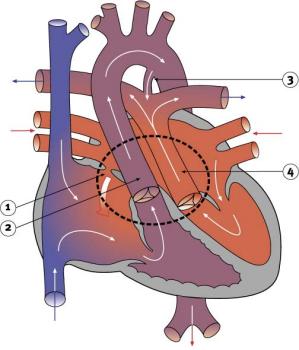
Transposition of the great arteriesIn transposition of the great arteries (also sometimes called transposition of the great vessels), the aorta and the pulmonary artery (the great arteries) are each connected to the wrong ventricles. The aorta is connected to the right ventricle, so that venous blood is carried to the body instead of to the lungs. The pulmonary artery is attached to the left ventricle, so that oxygen-rich blood is carried back to the lungs instead of to the body. Newborns with transposition survive only if they have one or more connections that let oxygen-rich blood reach the body. These connections may be in the form of a hole between the two ventricles, a patent foramen ovale or a patent ductus arteriosus. Most babies born with transposition are extremely blue soon after birth because their bodies are not receiving enough oxygenated blood. In order to immediately improve the body's oxygen supply, prostaglandin is given to keep the ductus arteriosus open. It is also possible to enlarge the patent foramen ovale by inflating a small balloon during a heart catheter procedure. This is known as a balloon atrial septostomy and allows more mixing between the right and left sides of the circulation. A common surgical procedure for treating transposition is known as an arterial switch. In this operation, the great arteries are disconnected from the ventricles to which they are attached at birth and are reconnected to the appropriate ventricles. The pulmonary artery is connected to the right ventricle, while the aorta is connected to the left ventricle. The coronary arteries are also re-implanted. Depending on the condition of the baby, this procedure may be done in the first few weeks after birth. The repair is more complicated if there is also a large VSD or other defects. An arterial switch operation is considered a high-risk procedure.
|
| Current | Home - Table of Contents - Chapter 2 - Transposition of the great arteries |
| Next | Double outlet right ventricle |
| Previous | Partial anomalous pulmonary venous drainage |
| Section 1 | Chapter 1 - Introduction to the Issues |
| Chapter 2 - Pediatric Cardiac Issues | |
| Chapter 3 - The Diagnosis of Pediatric Heart Defects and their Surgical Treatment | |
| Chapter 4 - The Health Sciences Centre | |
| Section 2 | Chapter 5 - Pediatric Cardiac Surgery in Winnipeg 1950-1993 |
| Chapter 6 - The Restart of Pediatric Cardiac Surgery in 1994 January 1, 1994 to May 17, 1994 |
|
| Chapter 7 - The Slowdown May 17 to September 1994 |
|
| Chapter 8 - Events Leading to the Suspension of the Program September 7, 1994 to December 23, 1994 |
|
| Chapter 9 - 1995 - The Aftermath of the Shutdown January to March, 1995 |
|
| Section 3 | Chapter 10 - Findings and Recommendations |
| Appendix 1 - Glossary of terms used in this report | |
| Appendix 2 - Parties to the Proceedings and counsel | |
| Appendix 3 - List of witnesses and dates of testimony | |
| Diagrams | |
| Tables | |