|
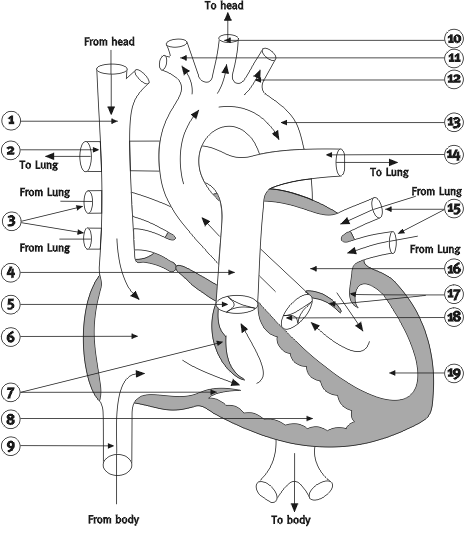
How a normal heart worksThe normal heart consists of four chambers: two upper atria (singular: atrium) and two lower ventricles. The atria are the collecting chambers of the heart, into which blood flows from the body and the lungs. The atria pump blood into the ventricles, although the force of the atrial pumping action is not as strong as that of the ventricles. These are the stronger pumping chambers, responsible for moving blood to the lungs and to the body. Of the two ventricles, the right ventricle is not as strong a pump as is the left ventricle. The right ventricle only pumps blood through the lungs or pulmonary circulation, which has a low resistance and therefore requires less pressure than the left ventricle, which pumps blood around the entire rest of the body. Thus although the amount of flow needs to be equal through the right and left sides of the heart, the pressures are quite different on the two sides. 
Blood flows from the body into the right atrium. That blood is 'blue' and is in need of oxygen, because oxygen has been removed from the blood by the body's organs and muscles in the course of blood flow. This blood is referred to as venous blood. It then passes from the right atrium into the right ventricle, which pumps the blood through the pulmonary arteries to the lungs where the blood picks up a fresh supply of oxygen. The oxygenated or 'red' blood then returns to the heart, flowing from the lungs through the pulmonary veins to the left atrium. The oxygenated blood next passes into the left ventricle, from where it is pumped through the aorta to the body. The heart has three major vessels that bring blood to and take blood from it. The superior vena cava (SVC) and the inferior vena cava (IVC) are the two largest venous vessels that connect to the right atrium. The SVC returns the blood from the head and upper body to the heart to be oxygenated, while the IVC does the same for the lower body. The aorta is the major arterial vessel that takes blood from the heart. The left ventricle pumps all the oxygenated blood through the aorta to the body. As blood flows through each of the four chambers of the heart, valves open and close in precise sequence. In the normally functioning heart, this allows blood to flow forward into the next chamber and prevents it from flowing backward. If a valve is defective, then blood can sometimes flow backward through it. When blood flows backwards through a valve, this is known as regurgitation. There are four valves in the heart. The mitral valve and the tricuspid valve separate the heart's upper and lower chambers. The tricuspid valve separates the right atrium from the right ventricle, while the mitral valve separates the left atrium from the left ventricle. These valves are known as the atrioventricular valves. The pulmonary valve separates the right ventricle from the pulmonary artery, which is the main blood vessel connecting the heart to the lungs. The aortic valve separates the left ventricle from the aorta. It is the closing of all four valves which produces the heart beat sound with its familiar 'lub -dub'.
When heard with a stethoscope, heart murmurs sound like soft whooshing noises that follow or replace the normal sounds of the heart's action. Murmurs may indicate that blood is leaking through an imperfectly closed valve and may signal the presence of a serious heart problem. The contractions of the heart are actually more complex than the simple squeezing of a bag. In effect, the heart contracts in rhythm, with the left ventricle contracting slightly ahead of the right ventricle. These contractions occur so closely together that one can think of them as one contraction, but that is not actually the case. The wall of the heart consists of three distinct layers-the epicardium (outer layer), the myocardium (middle layer) and the endocardium (inner layer). The myocardium actually causes the heart to contract, as its muscle fibres create a wringing movement that efficiently squeezes blood from the heart with each beat. The thickness of the myocardium varies according to the pressure generated to move blood to its destination. Thus the myocardium of the left ventricle is thicker than the myocardium of the right ventricle. The endocardium lines the cavities of the heart, covering the valves, small (papillary) muscles inside the heart and structures called chordae tendinae. (These are fibromuscular structures extending from the papillary muscles to the edge of the mitral and tricuspid valves.) The volume of blood expelled by the heart is referred to as the cardiac output. This is usually calculated on the basis of the volume of blood expelled with each contraction of the heart (or stroke volume), multiplied by the number of times that the heart beats each minute. Blood pressure is a measure of the force that results from the heart's pumping action. The highest (systolic) pressure occurs during contraction of the ventricles; the lowest (diastolic) pressure occurs during ventricular relaxation. When the blood pressure is too low, the patient is said to have hypotension. When blood pressure is too high, the patient is said to have hypertension. The heart is suspended in a membranous sac called the pericardium. The fibrous pericardium (the strong outer portion of the sac) is firmly attached to the diaphragm beneath it, to the membrane lining the chest on each side and to the sternum (or breast bone) in front. The heart is covered by the visceral pericardium or epicardium. Between the heart and the epicardium lies the pericardial cavity, which is normally filled with a very small amount of pericardial fluid. The pericardium can become irritated or inflamed following surgery. This condition is called pericarditis. This can be accompanied by an increase of fluid in the pericardial sac. If this fluid accumulates rapidly or in great amounts, the heart can become compressed by the fluid, a state called cardiac tamponade. The heart then has less ability to fill with blood and there is less cardiac output. Severe cardiac tamponade can cause a shock-like state that may be fatal. In such cases it is necessary to remove the fluid.
|
| Current | Home - Table of Contents - Chapter 2 - How a normal heart works |
| Next | Congenital Heart Disease |
| Previous | The Human Heart |
| Section 1 | Chapter 1 - Introduction to the Issues |
| Chapter 2 - Pediatric Cardiac Issues | |
| Chapter 3 - The Diagnosis of Pediatric Heart Defects and their Surgical Treatment | |
| Chapter 4 - The Health Sciences Centre | |
| Section 2 | Chapter 5 - Pediatric Cardiac Surgery in Winnipeg 1950-1993 |
| Chapter 6 - The Restart of Pediatric Cardiac Surgery in 1994 January 1, 1994 to May 17, 1994 |
|
| Chapter 7 - The Slowdown May 17 to September 1994 |
|
| Chapter 8 - Events Leading to the Suspension of the Program September 7, 1994 to December 23, 1994 |
|
| Chapter 9 - 1995 - The Aftermath of the Shutdown January to March, 1995 |
|
| Section 3 | Chapter 10 - Findings and Recommendations |
| Appendix 1 - Glossary of terms used in this report | |
| Appendix 2 - Parties to the Proceedings and counsel | |
| Appendix 3 - List of witnesses and dates of testimony | |
| Diagrams | |
| Tables | |